


Single-photon calcium imaging for interrogating the circuitry of the frontoparietal cognitive control network
Funding Program
BrainsCAN Accelerator Grant: Stimulus
Awarded: $60,944
Additional BrainsCAN Support
Western Faculty, Group or Institution
Department of Physiology & Pharmacology, Schulich School of Medicine & Dentistry
Keywords
Novel neuroscience/neuroimaging techniques, miniscopes
Related
none
Share this page
Background
Neuropsychiatric disorders are mental disorders that are attributable to diseases of the nervous system. A large number of these disorders show involvement of the 'frontoparietal cognitive control network', a network within the brain that enables complex problem-solving (by supporting diverse functions such as selective attention, decision-making and task switching).
However, we don't fully understand how these processes are represented by distributed populations of neurons in the brain, both in health and in disease. We believe the approach to understand this is to record large groups of individual neurons while a subject's brain is involved in tasks - while the subject is 'behaving'.
The Problem
Up to now, much of the empirical data used in developing our understanding, and more particularly our assumptions of the structures and neurons involved, has come from microarray electrophysiology. This method only samples neurons sparsely so alternatives have been developed. One is calcium imaging.
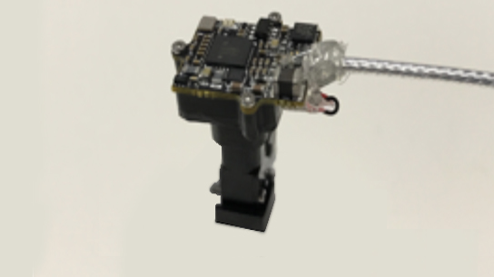
Figure 1 - A custom-built miniscope
Neuronal calcium concentration is strictly linked to electrical activity in a neuron, so we can monitor the electrical activity of hundreds of neurons by imaging the calcium concentrations. Calcium imaging can be achieved using a miniature fluorescence microscope, known as a miniscope. By incorporating a calcium indicator protein, this calcium concentration level can be detected as a fluorescence of the neuron.
This will enable us to use real imaging data of hundreds of neurons to build and test computational models of the frontoparietal network that are grounded in real neural circuit architecture.
The Project
We will use miniscopes to detect activities in the frontal eye field, a brain region responsible for voluntary eye movements and perception and awareness in the field of vision. The detection of neuronal activity with calcium imaging can then be compared with the visual stimuli and eye movements expected. If successful, this project will show the feasibility of calcium imaging using miniscopes in this way and open the door for future work to expand our understanding of frontoparietal cortical circuits.
Western ResearchersStefan Everling |
Figure credits
Figure 1 - the research team
© 2022 BrainsCAN Western University
This Summary is licensed under a Creative Commons Attribution-Noncommercial-No Derivative Works 4.0 License


